In Vivo Imaging System, FOBI
In Vivo Imaging System
Negotiable Min Order Quantity Unit
- Required Quantity
-
- Place of Origin
- South Korea
- Brand name
- In Vivo Imaging System, FOBI
- Payment Terms
- L/C,T/T
- Production method
- Available
- Shipping / Lead Time
- Negotiable / Negotiable
- Keyword
- drug delivery, oncology, fluorescence, cell tracking
- Category
- Other Clinical Analytical Instrument

CELLGENTEK Co., Ltd
- Verified Certificate
-
4


| Product name | In Vivo Imaging System, FOBI | Certification | CE |
|---|---|---|---|
| Category | Other Clinical Analytical Instrument | Ingredients | - |
| Keyword | drug delivery , oncology , fluorescence , cell tracking | Unit Size | 26.0 * 40.0 * 26.0 cm |
| Brand name | In Vivo Imaging System, FOBI | Unit Weigh | 9 kg |
| origin | South Korea | Stock | 10 |
| Supply type | Available | HS code | 902750 |
Product Information
Fluorescence In Vivo Imaging System
High Quality Image Data
Personal Imaging System
(Compact, Easy, Cost effective)
In Vivo, Ex Vivo and In Vitro
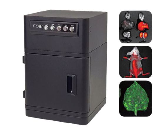
FOBI is a device that can image and analyze fluorescent signals from tissues and organisms. Images of various fluorescent proteins and dyes are taken using 4 channels consisting of Blue, Green, Red, and NIR. Using an optimized light source, filter, and color camera for macro-imaging, FOBI can obtain intuitive and high quality images. This configuration clearly distinguishes between background and signal without further analysis and is also available through a live window.
NEOimage program providing with FOBI analyzes fluorescence images easily by removing a background image effectively which is the biggest obstacle for fluorescence imaging and caused by autofluorescence and reflected light. In addition, the uniform light intensity of LED light makes it possible to measure certain quantity values. FOBI has a simple design, is easy to use, fast and reliable.
Feature
Intuitive Color Data
FOBI uses color sensors and optimized filters for the fluorescence signal through a live window without any special analysis. The live window allows you to identify the position and intensity of the fluorescence intuitively, and to get image data as it shown.

Fig. 1. Intuitive data
a. Image by FOBI’s color sensor.
b. Image by competitor’s mono sensor.
Fast
FOBI has a fast frame rate capable of recording videos. Due to the fast video speed, many samples can be processed quickly and observed, responded instantly.
Simple
FOBI is structured as simple and optimal for quick and easy installation. It is also easy to move, manage, and maintain.
Compact Size
The FOBI has a compact size (26 x 26 x 40 cm), so it is ideal for small spaces. Due to its convenient size and portability, it can be used for a wide variety of applications.
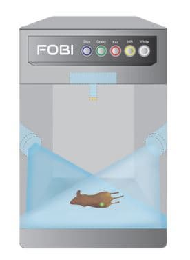
Fig. 2. Structure of FOBI
Easy to Use
Hardware and software are user-friendly. Mounting filters, controlling exposures, and capturing images are all simple and easy to use.
Multi function
It is possible to apply most fluorescence proteins and fluorescence materials from GFP to ICG by using four channels as Blue, Green, Red and NIR. Since more than one fluorescent substance can be imaged, various functions can be observed in one sample. For example, tumor and drug imaging can be performed in the same animal so that targeting and tumorization can be observed simultaneously. You can also merge bright images in order to localize the fluorescence within the animal.
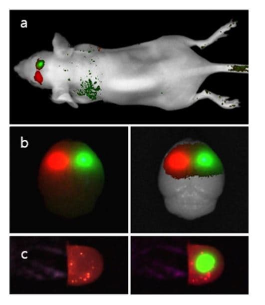
Fig. 3. Multi function imaging
a. In Vivo image of Tumor cell (green) and Stem cell (red) in the same brain.
b. Whole brain image after sacrificed.
c. Sliced brain image.
Application
Tumor Imaging
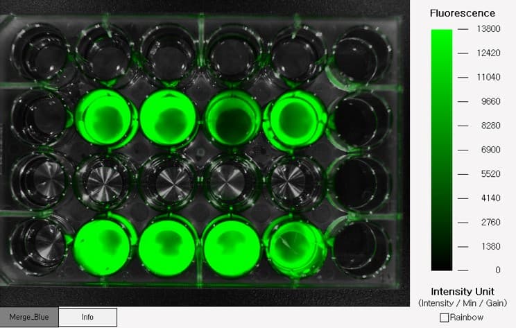
GFP stable cell line can be used to confirm tumorization. The created GFP stable cell line can be imaged In Vitro by using FOBI. By means of injecting GFP cells into subcutaneous tissues, cell proliferation can be imaged as fluorescence. In this way, it can obtain the images of metastasis to other tissues, in addition to quantifying and comparing tumor size.
Over the time, the signal strength of the fluorescence changes and the camera exposure time may vary accordingly. Because NEOimage analysis program can quantify the changes depending on imaging conditions like exposure time and gain, it can also compare and analyze the result of samples under the different condition.
Cell Tracking
Stem cells or immune cells with enhanced functions for various purposes can be imaged within the animal so as to ascertain their location and viability. Stem cells and immune cells are difficult to label with fluorescent genes. So, cells can be stained with fluorescent reagents in a variety of ways.
Stem cells and immune cells stained with a fluorescent reagent can be put into an animal using various methods such as intravenous injection, intraperitoneal injection, and subcutaneous injection. These cells can be located by using FOBI imaging. Cell survival can be determined by using quantitative analysis.
Plant
FOBI can image GFP labeled plant leaves. Plant leaves are difficult to obtain images of due to the strong autofluorescence of Chlorophyll. Chlorophyll’s autofluorescence can be removed and analyzed with GFP using a specific filter. The autofluorescence of chlorophyll itself can also be used as data. The degree of activity of chlorophyll can be confirmed by the intensity of the autofluorescence. In addition, images can be obtained from plant seeds and callus. Fluorescence imaging is possible with plants throughout their entire life cycle.
DDS(Drug Delivery System)
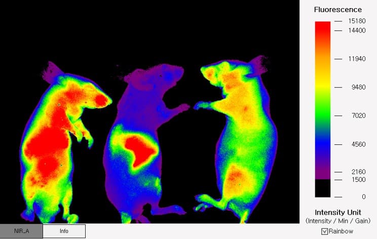
Drugs confirmed In Vitro can be injected into animals for experimental purposes. By taking images at certain intervals, you can check the movement and accumulation pattern of the drug in the living tissues of the animal.
The image of the drug confirmed In Vivo can be checked again Ex Vivo. Because the fluorescence is still expressed even after the animal is sacrificed, it is possible to quantify each tissue separately.
The resulting Ex Vivo data, together with the In Vivo data, can provide excellent evidence for an experiment.

Fig. 1. Animal imaging by FOBI
It is possible to apply most fluorescence proteins and fluorescence materials from GFP to ICG using four channels of Blue, Green, Red and NIR. Since more than one fluorescent substance can be imaged, different functions can be observed in one sample. For example, tumor imaging and drug imaging can be performed in the same animal, so targeting and tumorization can be observed simultaneously. You can also merge bright images in order to localization the fluorescence within the animal.

Fig. 2. Fluorescence imaging of various materials and methods
a. Fluorescence labeled chemicals in the Zebrafish. b. GFP cell in the 24well plate. c. Fluorescence labeling test. d. Ex Vivo imaging for drug delivery system. e. GFP expression leaf infected gene by virus vehicle. f. Auto-fluorescence from the chlorophyll. g. Gene expression on the leaf with marker gene. h. Gene transfected seed seperated by GFP imaging.
Specifications

Accessories
Product Type
There are two types of FOBI. One is a standard type that takes a picture with the door closed and outside light blocked.
The other is an open type with no doors and walls on the right and left. The open type FOBI can be used
when the sample size is large, such as rabbits and apes, or when recording a video of a surgical scene.

- Product Info Attached File
- Verified Certificate
-

B2B Trade
| Price (FOB) | Negotiable | transportation | Air Transportation,Express,Ocean Shipping |
|---|---|---|---|
| MOQ | Negotiable | Leadtime | Negotiable |
| Payment Options | L/C,T/T | Shipping time | Negotiable |

CELLGENTEK Co., Ltd
-
4


- President
- KIM HOE YUL
- Address
- 579 4 Yuseongdaero, Yuseong-gu, Daejeon, Korea
- Product Category
- Medical Analyzer
- Year Established
- 2003
- No. of Total Employees
- 1-50
- Company introduction
-
CELLGENTEK is a company that conducts studies and invents to create the most optimal bioimaging instrument. CELLGENTEK aims at the practicality of bioimaging instrument and the cost-efficiency at the same time. CELLGENTEK’s brilliant ideas and efforts would be served as a foundation for better biological research.
- Main Product
Related Products
Berry reusable nellcor oximax DB 9 Pins spo2 sensor
CRYSTE Multi purpose Centrifuge VARISPIN 4A
Compact Thermocycler DuxCycler
Orbital Shaker

IVD Point-of-Care Testing System LabGenius





 South Korea


