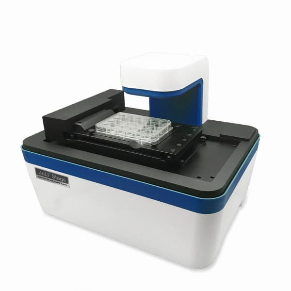
JuLI Stage
Real-time live cell imaging system
Negotiable Min Order Quantity Unit
- Required Quantity
-
- Place of Origin
- South Korea
- Payment Terms
- Others
- Production method
- OBM
- Shipping / Lead Time
- Negotiable / Negotiable
- Keyword
- incubator, real-time recording, cell imaging system, juli
- Category
- Other Equipment
NanoEnTek Inc.
- Recent Visit
- Jan 13, 2025
- Country / Year Established
-
 South Korea
/
2000
- Business type
- Manufacturer
- Verified Certificate
-
17


| Product name | JuLI Stage | Certification | - |
|---|---|---|---|
| Category | Other Equipment | Material | - |
| Keyword | incubator , real-time recording , cell imaging system , juli | Unit Size | - |
| Brand name | - | Unit Weigh | - |
| origin | South Korea | Stock | 0 |
| Supply type | OBM | HS code | - |
Product Information
KEY FEATURES
- Compact and compatible with a standard CO₂ incubator
- Fully automated X-Y-Z stage
- Multi-channel fluorescence imaging (GFP, RFP, DAPI and Bright field)
- Easy & powerful software
- Take and analyze image in real-time


- Product Info Attached File
B2B Trade
| Price (FOB) | Negotiable | transportation | Negotiation Other |
|---|---|---|---|
| MOQ | Negotiable | Leadtime | Negotiable |
| Payment Options | Others | Shipping time | Negotiable |
- President
- Chanil Chung
- Address
- Guro-gu, Guro-dong,235-2, Guro-gu, Seoul, Korea
- Product Category
- Medical Devices
- Year Established
- 2000
- No. of Total Employees
- 101-500
- Company introduction
-
- Main Markets
-
 Germany
 Italy
Italy
 Japan
Japan
 Taiwan
 U. Kingdom
- Main Product
Related Products
LIO FLEX Extreme Cold
SUNSHINE MULTI COLORED FLAME SYSTEM
Portable, Endoscopic visual system_QVION
Food Waste Microbial Decomposer(TB-99KL)
_2.jpg)
Cable and Wire harness tester (MHT-610 / 705)


